Skin tears are common injuries to the skin that affect various patient populations, especially the aging population. According to the Centers for Disease Control and Prevention (CDC), older adults will make up nearly 25% of the U.S. population by 2060. And as this group continues to age, the risk for chronic diseases and susceptibility to this wound type increases.
The prevalence of skin tears continues to rise, with some studies estimating as many as 1.5 million occurrences of skin tears each year in the U.S. Another study reports higher skin tear incidences in long-term care facilities, with 2.1% among male residents and 4.6% among female residents.
In a session at the 2023 Wild on Wounds conference, wound care clinicians learned more about skin tears, understanding the risks, complications, and treatment methods. Here, we take a closer look at these traumatic wounds as well as some key points from this session, including classification systems and ways to prevent them.
What are skin tears?
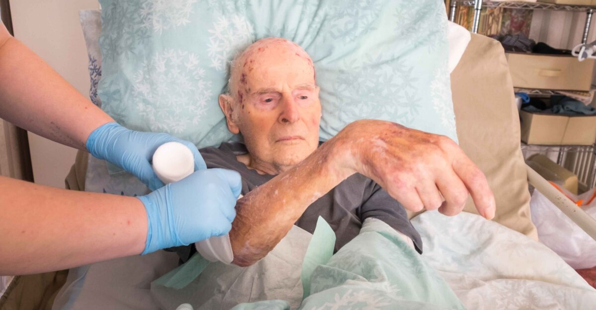
Skin tears are traumatic wounds, resulting from forces that include friction, shearing, blunt force trauma, falls, equipment, poor handling, and medical adhesive-related skin injury (MARSI). These mechanical forces cause the layers of skin to separate.
These wounds can be common within different patient populations, including those who are very young or critically ill. But skin tears are most prevalent in the aging population. This is due to skin losing elasticity, thinning of the epidermis and dermis over time, and loss of fat. Due to these age-related skin changes, blood vessels are more likely to be damaged, causing bruising and increased bleeding with even mild trauma to the skin.
Classifying skin tears
Founded in 2011, the International Skin Tear Advisory Panel (ISTAP) is the newest method of classifying skin tears. This classification system separates tears into three levels that include the following:
- Type 1: No Skin Loss — This type of skin tear includes linear tears or skin flaps that can be repositioned to cover the wound bed.
- Type 2: Partial Flap Loss — This type of tear has tissue loss extensive enough that only part of the flap can be repositioned over the wound bed.
- Type 3: Total Flap Loss — These skin tears have no tissue flap attached. There is only an open wound bed.
Prior to the creation of the ISTAP classification system, wound care nurses and other clinicians used the Skin Tear Classification System (STAR) or the Payne-Martin system, which provided similar categories for skin tears.
Treatment methods
Early treatment of skin tears improves patient outcomes and can prevent unnecessary tissue loss. When implementing treatment, the goal is to salvage viable tissue, heal the wound bed, and protect the periwound skin from further trauma.
The first step in treatment of an acute skin tear is to control bleeding and gently cleanse the wound once bleeding has subsided. Cleanse the wound with normal saline or clean tap water. Then attempt to salvage any attached skin flaps by gently repositioning them over the wound bed. If the skin flap is not viable, a medical provider may need to debride the necrotic tissue. Dressing selection for skin tears should consist of dressings that control drainage, keep the wound bed warm and moist, and protect the wound/periwound skin from further trauma — especially during dressing removal.
In the past, transparent film dressings and hydrocolloids were routinely used, but these types of dressings typically have strong adhesives and can pose challenges with these wound types. Silicone foam dressings are a good choice as they provide protection, are easier on the skin during removal, and can stay in place up to seven days.
Appropriate-sized dressings should be used, and skin barrier should be applied wherever the dressing will touch the skin. Removing the dressing “low and slow” will help prevent epidermal stripping.
Complications, risk factors, and prevention
As with any break in our protective skin layers, skin tears are susceptible to infection. An untreated infection can progress to surrounding tissues. And if infection continues without treatment, systemic infection and sepsis can occur.
Along with complications from infection, risk factors can increase the chances of these wounds occurring or complicate existing ones. The most common risk factors include:
- Aging
- Chronic medical conditions
- Certain medications, such as anticoagulants, aspirin, steroids, and diuretics
- Poor mobility
- Poor vision
- Dry skin
- Dehydration
- Poor nutrition and hydration
The key to prevention is early intervention. This means educating patients on ways to best prevent skin tears, which include:
- Maintaining adequate hydration and nutrition. Dehydrated or unhealthy skin is more prone to injury.
- Evaluating home environments and making modifications to avoid unnecessary bumps, trips, and falls. This means educating patients to keep walkways clear and avoid the use of rugs that can move and slip. Encourage the use of a light at night as well as the use of assistive devices, such as walkers and canes when needed.
- Moisturizing the skin, especially over areas that have had tissue injury in the past. Avoid over drying the skin with harsh soaps and perfumed lotions. Check that topical products do not contain alcohol, as this can dry the skin out further.
- Avoiding the use of non-silicone adhesive bandages and medical tapes.
- Using pH-Balanced products and avoiding excessive bathing which can over-dry the skin.
When to see a medical professional
Skin tears often heal with good care. But like other wounds, they are not immune to become more serious.
For instance, a skin tear on the lower legs that does not heal within three weeks is at risk to become a complex wound. And if a patient has comorbidities such as diabetes, autoimmune disorders, lymphedema, and peripheral vascular disease, this can create even more risk.
While every wound is different, each patient should know when to seek out medical care. If patients experience any of the following, they should see a medical professional:
- Signs of infection, including redness, significant pain, fever, nausea, chills, swelling, warmth, increased drainage, and/or odor
- A large or deep area of tissue loss.
- Uncontrolled bleeding
- Ongoing or increasing pain
- Wound not showing improvement after two weeks
Skin tears have been described as “not a big deal.” However, these injuries increase healthcare costs and impact quality of life. They can cause psychosocial problems such as pain, sleep disturbance, and mobility problems. Draining or large wounds can lead to social isolation. For these reasons, providing good care to even minor skin tears can be transformative to the lives of your patients.
Take our engaging, evidence-based Wound Care Certification Courses for nurses, registered dietitians, physical therapists, and more professionals. Choose the format that suits you and get access to tools to help you ace your exam.
Learn MoreWhat do you think?