
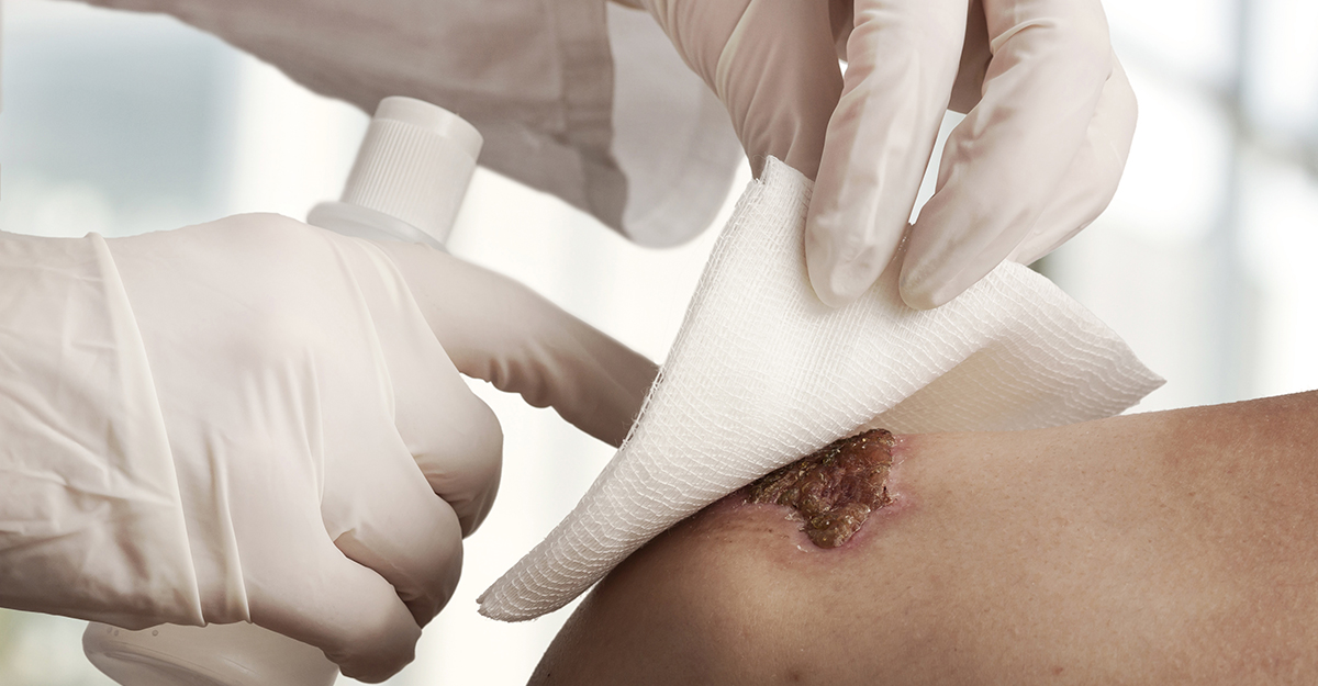
Every wound care clinician has had that moment. You remove a dressing, look at the wound, and think: Wait, what am I seeing?
Some wound findings naturally demand attention. The tissue may appear dark, wetter than expected, raised, discolored, or simply different from what was documented during the previous assessment.
But a change in appearance does not necessarily indicate a complication.
A wound’s appearance is only one piece of the clinical picture. Etiology, wound-bed tissue, drainage, surrounding skin, pain, perfusion, infection risk, and changes over time are all important to a comprehensive wound assessment.
A chronic wound assessment review published in American Family Physician recommends systematically documenting a chronic wound’s location, size, depth, drainage, and tissue type. It also emphasizes identifying the wound’s underlying cause rather than focusing only on its appearance.
What these wound findings can reveal
Here are five wound findings that deserve a closer look and what they can reveal during a comprehensive assessment.
1. Eschar: When black doesn’t tell the whole story
Dark or black tissue in a wound can immediately grab your attention.
Eschar is dead tissue that may appear brown or black and can be firmly attached to the wound bed. But recognizing eschar is only the beginning of the assessment.
As WCEI explains in Eschar vs Slough: Know the Difference, you should look beyond color when distinguishing wound tissue. Texture, moisture level, adherence, wound etiology, and perfusion status can all help provide a more complete picture. However, the appearance of black tissue should not automatically dictate the intervention. National Pressure Injury Advisory Panel guidance states that stable eschar — dry, adherent, and intact, without erythema or fluctuance — on the heel or an ischemic limb should not be softened or removed.
So, before taking the next step, ask yourself some additional clarifying questions: Where is the wound located? Is the tissue dry or moist? Is it stable? What is the suspected wound etiology? Is perfusion adequate? Are there signs of infection or instability?
Clinical takeaway: Avoid making a treatment decision based on color alone. Let the complete assessment guide whether to protect and monitor the eschar or seek further evaluation for possible removal.
2. Slough: The yellow finding that deserves context
Yellow in a wound bed may represent slough, exudate, dressing residue, or another structure.
Yellow color in a wound bed should not automatically be associated with pus.
Slough is material containing devitalized tissue and inflammatory debris that may appear yellow, white, tan, or gray and can vary in consistency and adherence. The International Wound Infection Institute (IWII) identifies purulent drainage as an overt sign of local wound infection, but it may appear in other colors besides yellow.
That distinction matters.
The Cleveland Clinic’s overview of purulent drainage describes purulent drainage as thick or milky fluid that can appear white, yellow, green, pink, or brown. It may also have an unpleasant odor and is associated with infection.
When yellow material is present, determine whether you’re looking at tissue within the wound bed or drainage coming from the wound.
Clinical takeaway: When yellow material is present, first determine whether it is wound-bed tissue or drainage. Then assess the wound bed, drainage, pain, warmth, swelling, odor, and periwound skin, and compare the findings with the prior assessment. Describe the finding before interpreting it. Color is a clue, not a diagnosis.

3. Hypergranulation: When there’s too much of a good thing
Granulation tissue is generally a welcome sight in a healing wound. But an excessive amount can become a concern.
Hypergranulation is excess granulation tissue that rises above the surrounding skin and can interfere with epithelial tissue advancement. The tissue may look raised, moist, red, or friable, and its appearance can make clinicians wonder whether something has gone wrong.
A review on granulation tissue available through the National Library of Medicine identifies infection, excess inflammation, foreign material, and physical irritation or friction among possible contributors to hypergranulation.
The raised tissue is only part of the picture. The next question is why it may be happening.
Clinical takeaway: Hypergranulation is a finding that warrants further assessment rather than an assumption that it represents normal healing. Look for potential contributing factors rather than focusing only on the tissue’s raised appearance.
4. Unexpected drainage: When the dressing tells a story
Sometimes the biggest surprise isn’t the wound bed. It’s the dressing you just removed.
Maybe the dressing is considerably more saturated than it was at the previous dressing change. Perhaps the fluid has changed color or consistency. Or maybe drainage that was previously minimal is suddenly much more noticeable.
Not all drainage is abnormal. Some wound drainage is expected during healing, but its characteristics and changes over time can provide clinical context.
For example, Cleveland Clinic’s medically reviewed resource on serous drainage explains that small amounts of clear-to-yellow serous fluid can be part of normal wound healing. Large amounts of fluid or a change toward thick, pus-like drainage, however, warrant further evaluation.
For clinicians, the trend matters.
Assess the exudate amount, color, consistency, and odor, along with leakage and dressing saturation. Interpret these findings in relation to the dressing type, its fluid-handling capacity, and its wear time. Compare the findings with previous assessments, recognizing that a new concern may require evaluation even without an established baseline.
Clinical takeaway: Instead of asking only, “Does this drainage look concerning?” ask, “Is this different from what we’ve been seeing?”
A change in drainage may be more informative than a single observation.
5. Periwound changes: When the clue is outside the wound
The wound bed tends to get most of the attention. Sometimes the bigger clue is sitting right beside it.
Changes in the periwound skin may include maceration, edema, induration, warmth, tenderness, dryness, discoloration, or loss of skin integrity. These findings can raise questions about moisture management, inflammation, infection, pressure, medical adhesive-related skin injuries, or other factors affecting the wound environment.
The IWII’s 2026 consensus document identifies local warmth, swelling, changes in skin color, and new or increasing pain among findings that may indicate wound infection.
Of course, you should interpret those findings alongside exudate, wound progression, and the patient’s overall condition. Assessment should also account for baseline skin tone. In darker skin, inflammation may appear as an area that is darker than the patient’s normal skin tone or has blue or violet discoloration rather than obvious redness.
Clinical takeaway: Expand your field of view. Examine the wound edges and surrounding skin and compare what you’re seeing with the patient’s baseline and previous assessments.
Turn a double take into a closer assessment
Unexpected findings call for a deliberate assessment and appropriate response. This is the time to pause and take a closer look.
When you notice an unexpected change, resist the urge to jump directly from appearance to conclusion. Identify what you’re seeing, compare it with previous findings, consider the wound’s etiology, and assess the whole patient, not just the wound.
Clinical takeaway: What’s different? What else am I seeing? What could be contributing to this change? These questions can turn an unexpected finding into useful clinical information.
Findings suggesting spreading infection, systemic illness, or compromised perfusion require timely escalation.
Related reading: Learn more about recognizing and managing moisture-related periwound changes in Wound Care Nurse’s Guide to MASD Wounds.
See where wound care education can take you. Save $100 on eligible WCEI courses from October 25 through October 31, 2026, with code SPOOKY100.
Explore courses

What do you think?