
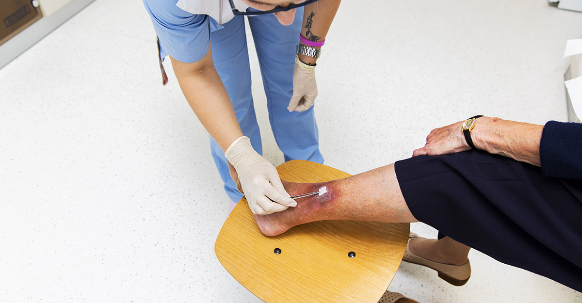
Cellulitis is a common skin and soft tissue infection that can be difficult to identify in wound care settings. Early recognition and intervention can help prevent complications, reduce hospitalizations, and support better outcomes for patients with wounds and skin injuries.
The Centers for Disease Control and Prevention (CDC) identifies cellulitis as a bacterial infection involving the deeper layers of the dermis and subcutaneous tissues that requires prompt medical evaluation and treatment.
Looking beyond redness
One of the biggest challenges in wound care is that cellulitis can resemble several other conditions. Venous stasis dermatitis, lipodermatosclerosis, contact dermatitis, and lymphedema can all cause erythema, edema, and changes in skin appearance.
However, cellulitis typically presents with several hallmark findings that help differentiate it from other conditions. Patients often report pain and tenderness in the affected area, along with localized warmth, swelling, and erythematous skin that may appear taut or shiny. According to the CDC, these findings are especially common in the lower extremities
An important clue is laterality. Lower-extremity cellulitis is typically unilateral. Bilateral lower leg erythema, particularly in the absence of tenderness, warmth, fever, or other signs of infection, should prompt consideration of alternative diagnoses.
Systemic symptoms can provide another important clue. Fever, chills, malaise, and fatigue should raise suspicion for an active infection rather than a purely inflammatory process. However, the absence of fever does not rule out cellulitis. Some patients may also develop lymphangitis, which appears as red streaking extending proximally from the infected area and may indicate that the infection is spreading through the lymphatic system.
You should also remember that erythema may be less apparent in darker skin tones. Assessment of warmth, tenderness, edema, skin texture, discoloration, and change from the patient’s baseline becomes particularly important.
How does cellulitis develop?
Cellulitis develops when bacteria gain access through a break in the skin barrier. While this seems straightforward, the entry point is not always obvious.
For wound care patients, risk factors may often present long before cellulitis develops. Venous leg ulcers, diabetic foot ulcers, pressure injuries, skin tears, surgical incisions, and traumatic wounds all create opportunities for bacteria to enter the skin. Even seemingly minor issues such as cracked skin from athlete’s foot (tinea pedis) can serve as a portal of entry.
The most common pathogens are beta-hemolytic streptococci and Staphylococcus aureus. Although many clinicians automatically think of MRSA when discussing skin infections, streptococci remain the primary cause of nonpurulent cellulitis. Treatment decisions should reflect the patient’s presentation, risk factors, and local antimicrobial stewardship guidelines.
Certain patient populations face a higher risk of developing cellulitis, including those with:
- Chronic edema
- Lymphedema
- Venous insufficiency
- Obesity
- Diabetes
- Impaired immune function
- Recurrent skin infections
These conditions can also increase recurrence risk, so prevention should be part of long-term management rather than an afterthought.

Is it colonization, cellulitis, or wound infection?
In wound care, distinguishing between colonization, cellulitis, and local wound infection is essential because each finding has different implications for assessment and treatment.
Chronic wounds are frequently colonized by microorganisms, and the presence of bacteria in a wound does not itself indicate clinical wound infection.
Cellulitis involves infection of the deeper dermis and subcutaneous tissue and typically presents unilaterally with spreading warmth, tenderness, erythema, and edema.
Local wound infection is changes in wound characteristics centered within the wound and the immediate wound border.
Note: Clinical findings, not just culture results alone, should guide the diagnosis and management of infection.
When animal bites and wounds change the picture
Not every case of cellulitis follows the typical clinical pattern.
Animal bites, human bites, puncture injuries, immersion injuries, and heavily contaminated wounds may introduce a broader range of microorganisms. In these situations, you should consider the possibility of polymicrobial infection rather than infection from a single organism.
The CDC notes that cellulitis associated with animal bites or atypical pathogens may warrant culture collection and more targeted antimicrobial management.
This is particularly relevant in wound care, where chronic wounds can contain multiple organisms and where infection management often requires a more comprehensive assessment than standard community-acquired cellulitis.
Red flags every clinician should recognize
Most cases of cellulitis respond well to treatment when identified early. However, you should know when the situation requires urgent escalation.
Immediate medical evaluation is warranted when erythema spreads rapidly, systemic symptoms develop, or the patient becomes acutely ill. The CDC specifically recommends prompt evaluation when fever, chills, or quickly expanding erythema occur.
Additional warning signs include:
- Rapid progression despite treatment
- Severe or worsening pain, out of proportion to the clinical examination
- Blister formation
- Purulent drainage
- Increasing edema
- Signs of sepsis
- Hypotension
- Mental status changes
- Crepitus or concern for abscess
- Clinical findings associated with necrotizing soft tissue infection
When appropriate, outlining and dating the outer margin of erythema with a skin-safe marker can provide an objective reference for monitoring progression or response to treatment.
Failure to begin improving after 48 to 72 hours of appropriate antibiotic therapy should also prompt reassessment. Persistent symptoms may indicate resistant organisms, an alternative diagnosis, or a more complex underlying condition. It is important to recognize that skin discoloration and erythema may take longer to completely resolve even when treatment is effective.
Treatment is more than antibiotics
Antibiotics remain the foundation of cellulitis treatment, but successful outcomes often depend on addressing the risk factors that allowed the infection to develop.
Current clinical guidance recommends prompt antibiotic therapy targeting likely pathogens, with adjustments made based on clinical response and culture results when available.
Supportive measures are equally important. Elevating the affected extremity can help reduce edema, improve lymphatic drainage, and decrease patient discomfort. Merck Manual Professional recommends immobilization and elevation of the affected area as part of cellulitis management.
Clinicians should also evaluate and manage underlying contributors, including uncontrolled edema, fungal infections, poorly fitting footwear, hyperglycemia, and untreated wounds. Addressing these factors not only supports healing but may help reduce the risk of recurrence.
Final thoughts
For wound care professionals, cellulitis is more than localized redness. It is a potentially serious bacterial infection that can progress quickly in vulnerable patients. Effective management requires recognizing symptoms such as pain, tenderness, swelling, fever, chills, and red streaking; identifying vulnerable patients; knowing when to escalate care; and addressing underlying factors that contribute to infection.
A careful assessment, prompt treatment, and attention to the whole patient can make the difference between a manageable infection and a preventable hospitalization.
Continue your learning journey with WCEI courses.
Explore courses

What do you think?