Key Takeaways
Wound debridement, the removal of nonviable tissue and microorganisms from wound beds, is crucial for effective wound healing and can be conducted through various methods including sharp, mechanical, autolytic, enzymatic, and biological techniques. The choice of debridement method depends on factors such as wound type, practitioner expertise, and patient tolerance, though each method is aimed at optimizing the wound bed for healing. Clear communication with patients about the debridement process and outcomes is essential to manage expectations and improve wound care experiences.
According to the National Library of Medicine, wound debridement, or the removal of nonviable tissue, biofilm, and bioburden from the wound bed, is an essential part of standard wound treatment.
Bioburden consists of microorganisms on the surface of the wound bed, while biofilm is a substance created by the presence of bacteria, microbes, and cellular debris. The removal of these tissues optimizes the wound bed for ongoing granulation of healthy tissue and promotes epithelialization.
Tissues like these act as barriers to wound healing, and chronic wounds will be unable to heal if these tissues are present — making debridement an essential part of the healing process.
Debridement may be performed in a practitioner’s office, an outpatient wound care clinic, or at the bedside during an inpatient stay. More extensive sharp and/or excisional debridement is performed by either a general surgeon, plastic surgeon, orthopedic surgeon, or surgical podiatrist.
Operating room debridement is usually required for larger wounds with extensive amounts of necrotic tissue. This often includes larger pressure injuries over the sacrum, buttock, and trochanter. It may also be necessary for wounds that have become infected in the deeper tissues and structures such as the tendon, muscle, fascia, or bone. This is often necessary for pressure injuries and diabetic wounds of the lower extremity.
The choice of one type of debridement over another depends on the type of wound, characteristics of the wound, practitioner training, product availability, and patient’s tolerance level for debridement.
Types of debridement
Here are the most common types of wound debridement:
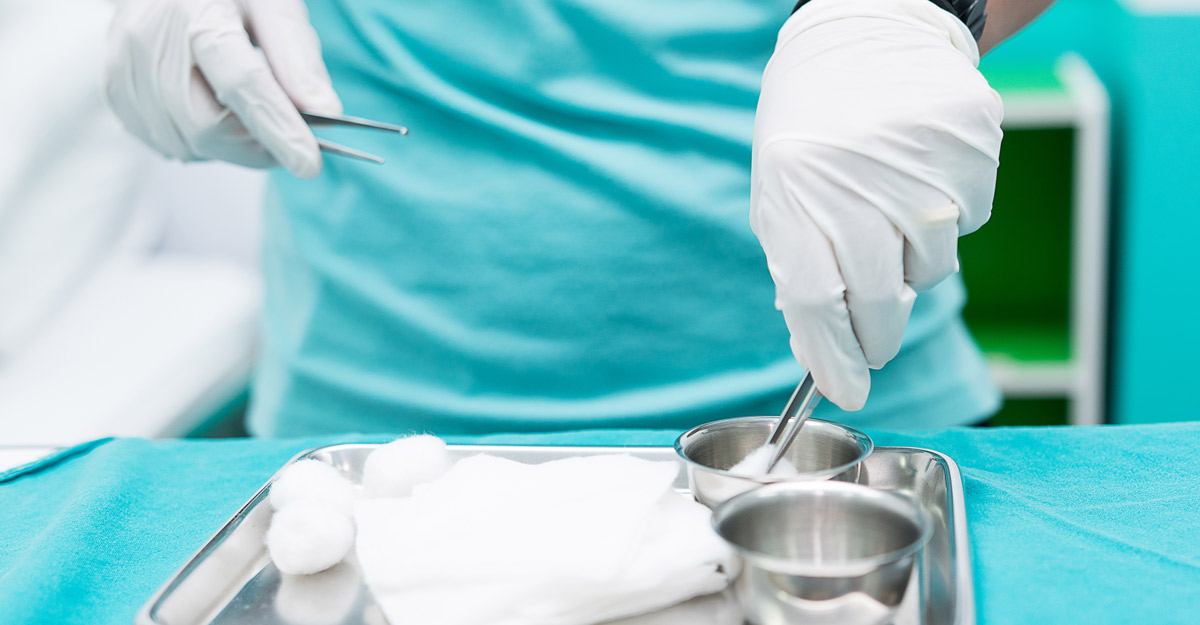
Sharp debridement
A common type of debridement within a clinical setting, sharp debridement is highly effective and the quickest way to remove devitalized tissue. A topical anesthetic is applied to the wound prior to the procedure, and an informed consent should be obtained prior to each sharp debridement. Several types of instruments may be used such as: curette, blade, forceps, and sterile scissor. A debridement of this type may extend into the subcutaneous tissue causing bleeding. This is normally managed by a brief period of pressure applied to the area. In some cases, it may be necessary to control bleeding by use of cauterization to the site. Patients on an oral blood thinner will be at increased risk of bleeding during this type of debridement. A review of medications prior to the procedure is important.
Mechanical debridement
Mechanical debridement removes the necrotic tissue from the wound bed using force. This type of debridement is nonselective meaning it will remove viable tissue along with the nonviable tissue. Methods of mechanical debridement include wet to dry dressings, wound irrigation and “scrubbing” of the wound bed with gauze and saline. Care should be taken as this type of debridement is usually painful for the patient.
Autolytic debridement
Autolytic debridement is one of the slower types of debridement but is also usually a pain free method. Autolytic debridement uses the body and specifically chosen wound dressings to create a “greenhouse effect” to the devitalized tissue — over time, creating a warm, moist environment will cause the necrotic tissue to break down, loosen, and become non adherent. The most common dressings used for autolytic debridement are films, gels, and hydrocolloids. This is a lower cost debridement that is also a good choice for patients who cannot tolerate a mechanical or sharp debridement.
Enzymatic debridement
Enzymatic debridement is another, usually painless, method. A collagenase ointment is applied to the wound bed where enzymes in the ointment break down the collagen in nonviable tissue. The collagenase ointment is selective, meaning it will break down only necrotic tissue such as slough and eschar. This method of debridement is normally faster than autolytic debridement but may be cost prohibitive for some patients.
Biologic debridement
Biological debridement is a centuries old treatment using maggots and leeches to improve wound bed tissue and viability. Maggots use their digestive enzymes to dissolve nonviable tissue leaving viable tissue unharmed. Leech therapy is useful to decongest tissues and improve vascularization at a specific site. This type of debridement is the least common in modern, conventional wound care as it requires lab grown maggots and leeches.
Debridement is beneficial for most types of wounds, although, an arterial ulcer should not be debrided. Debridement of an arterial ulcer may lead to further ischemia which can cause the condition of the ulcer to worsen. When eschar is present on toes, ankles, lower legs, or fingers, noninvasive arterial studies and a referral to a vascular surgeon are necessary to determine arterial perfusion prior to any type of debridement.
Likewise, debridement of dry eschar over a pressure injury to the heel is normally avoided. This is due to the dry eschar acting as the body’s biologic covering to protect the area. Over time, and with appropriate offloading, the eschar will begin to lift away. As the edges lift, the clinician may trim away the loose ends leaving the remaining adherent eschar alone. If, at any time, this adherent eschar becomes soft, wet, or spongy, debridement is warranted, as this can be a sign of infection.

Wound debridement considerations
No matter which type of debridement is chosen, it is likely your patient will need ongoing, serial debridement to achieve a healthy granulating wound bed free of nonviable tissue and low bioburden levels.
A wound that has undergone this treatment, especially a more extensive debridement, may look significantly different than they did prior to the debridement. An extensive debridement can temporarily make the wound measure larger and may change the way the wound bed looks. This new appearance may show a deeper, more cavernous wound bed that appears “worse” than before the debridement.
For this reason, prior to any type of debridement, clinicians should have an educational conversation that informs the patient and their support persons of what to expect from the procedure.
This can clear up any misperceptions the patient and family may have about the wound appearance following the procedure. Open communication and empowering the patient with knowledge can make the ongoing process of wound care and management easier on the patient, their support persons and families, as well as those who care for them.
Elevate your expertise with education from WCEI!
Learn MoreWhat do you think?