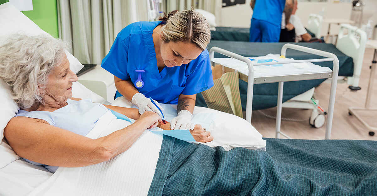
When teaching, I often get the question “What’s my role in the different stages of wound healing?”
To address this common question, I thought a review of basic wound physiology and the clinicians’ role during each of the stages of wound healing (aka phases of wound healing) would be helpful.
We know that the four phases of wound healing are driven by a mixture of chemical stimuli (growth factors and cytokines). Any diminished or excessive levels of these different chemicals can have a negative impact on the wound healing process. The phases are continuous and overlap each other to some extent. However, they must occur in a particular sequence to result in a healed wound.
No phase can be skipped, and each phase can last for a different length of time. However, the length of time can be influenced by what we do as clinicians. Let’s take a look at each phase and our role within them.
Hemostasis Phase
This phase occurs immediately upon tissue injury and initiates the entire wound healing process. The injured area will be filled with blood, histamine, serotonin, and prostaglandins.
Due to the damaged endothelial lining of the vessels, platelets will be activated to stop the bleeding by changing shape, increasing adhesiveness, clumping in the hole of the vascular wall, and releasing growth factors.
This causes the formation of thrombin, which stimulates the formation of a fibrin mesh to strengthen and secure the platelet “plug,” or clot, from being washed away by the flow of blood.
The clot, platelets, and surrounding wound tissue, release a multitude of pro-inflammatory cytokines and growth factors to direct the wound into the next phase of healing. In the event of a traumatic injury, clinicians can intervene with pressure, clotting/hemostatic agents, and cauterization (if necessary) to stop the bleeding.
The challenge with many chronic wounds is the lack of recent trauma to initiate the healing process properly. This results in senescence of important cells needed to repair the tissue and diminished production of growth factors.
Unfortunately, there is not a lot that clinicians can do to influence what happens during this phase.
Inflammatory/Defensive Phase
This phase is often referred to as the “clean up” phase in which the goal is to remove the debris and microbial contaminants in the wound. Neutrophils are the first to arrive and begin this process by destroying the microorganisms within the wound. However, their lifespan is quite short and they rapidly decrease in numbers after three days.
Monocytes, which differentiate into macrophages, arrive shortly after the neutrophils and become the “long term” clean-up crew. They consume the cellular debris, microorganisms, and foreign substances. Macrophages will eventually secrete growth factors and cytokines to trigger cells of repair like fibroblasts. The body also releases fibrinolytic and proteolytic enzymes to break down necrotic tissue.
In the case of an acute wound, there is very little debris. Therefore the body moves through this phase fairly quickly. However, with chronic wounds there is typically a significant amount of necrosis and bioburden issues that the body alone cannot manage in an efficient manner on its own.
This is the phase, or stage of wound healing, that clinicians can have an immediate impact on the speed in which the wound moves through it. Remember, the ideal wound for healing has a clean, viable wound base.
We can actively debride wounds with sharps, enzymes, chemicals, maggots, and a multitude of mechanical debridement options to expedite how quickly we achieve that clean, viable base. The goal should be to accomplish that as quickly as possible.
The inflammatory phase does not “heal” the wound, and since the ultimate goal is healing, we should spend the least amount of time we can cleaning the wound. Far too often we see approaches that do not expedite debridement of the wound in the most effective and efficient manner.
Proliferative Phase
This phase often overlaps with the inflammatory phase, especially in chronic wounds, and is the phase in which wounds “heal” or close. It is during this phase that the body produces granulation tissue to fill in the defect, contracts the edges to close the opening, and finally covers the wound with new epithelial tissue. This obviously occurs more efficiently when the wound is completely free of necrosis, debris, and high bioburdens.
The most dominant cell is the fibroblast, which synthesizes the collagen fibers that make up the granulation tissue (connective tissue) that replaces the injured tissues. It also plays a role in forming elastin, fibronectin, glycosaminoglycans, and proteases needed for remodeling, as well as keratinocytes for re-epithelialization.
Another important process that needs to occur is angiogenesis or the formation of new blood vessels in the form of capillary beds. Collagen synthesis in conjunction with angiogenesis results in the granulation tissue. Upon completion of this phase, the wound is closed with epithelial tissue.
The role of the clinician should be one of “laissez-faire” or “less is more.” This is to say that the body has to do all the heavy lifting during this phase, and our role should be more supportive by:
Reducing the frequency of dressing changes
Managing the bioburden to prevent infection and biofilm
Ensuring adequate nutrition and a moist wound environment
Continuing to treat/correct the underlying etiology
The key is less involvement in the wound (unless exudate management continues to be an issue), and more involvement in treating the entire patient to optimize healing like ensuring adequate nutrition and management of their underlying diseases processes that affect healing. Clinicians can also consider adjunctive modalities that can help enhance the process.
Maturation/Remodeling Phase
The fourth and final phase primarily occurs after the wound is closed. This phase can last a year or longer depending on the size of the wound and the amount scar tissue. Some wound contraction may occur, and fibroblasts will decline in number. Capillary density will diminish leading to the change in color of the scar from pink to white.
The initial Type III collagen will be converted into Type I collagen to increase the strength and structure of the scar tissue. In the early weeks, the tensile strength is only at 20%, and with enough time, will eventually achieve about 80% tensile strength of the original tissue. This of course means that the wound will be at higher risk for breakdown indefinitely.
As a clinician, the primary role is to protect the newly healed tissue from further damage and continue to support the patient’s nutritional needs. Modalities such as ultrasound can increase the tensile strength of the scar tissue quicker, as well as scar management techniques such as compression or soft tissue massage, to reduce keloid formation and improve scar smoothness can be introduced.
So those are the phases or stages of wound healing in a nutshell. Until next time. Heal on!
Take our engaging, evidence-based Wound Care Certification Courses for nurse, registered dietitians, physical therapists, and more professionals. Choose the format that suits you and get access to tools to help you ace your exam.
What do you think?


