Key Takeaways
Living with necrotic tissue poses significant challenges for patients, requiring evidence-based treatments from skilled clinicians to prevent life-threatening complications and improve quality of life. Necrotic tissue results from prolonged ischemia and can manifest in various forms, with treatment focusing on addressing the underlying causes and utilizing techniques such as debridement and specialized care protocols. Understanding and effectively managing necrotic tissue is crucial for patient well-being, highlighting the importance of comprehensive wound care education and intervention.
Living with necrotic tissue is challenging for patients and requires evidence-based treatments from skilled wound care nurses and clinicians to achieve better outcomes.
Research has shown the serious, life-threatening complications that arise when an infection from necrotic tissue is present.
The quality of life for patients living with necrotic wounds can be severely impacted. Among the more serious complications, patients also face challenges with mobility, pain, and the increased risk of reoccurring treatment.
Understanding the causes and signs of necrotic tissue and finding the most effective treatment can be life-changing for these patients. We spoke with two wound care experts about what necrotic tissue is, how to spot it, and the best course of action.
If you’re looking to gain a more in-depth knowledge of how to provide skin & wound care, our Skin and Wound Management courses can help.
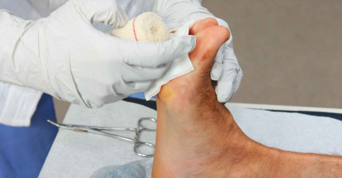
What is necrotic tissue and skin necrosis?
Skin Necrosis
The first step in understanding necrotic tissue is to understand necrosis. The term necrosis stems from the Greek work nekros, which means death.
“Necrosis is a loose term, and it can appear in two ways — under a microscope and viewed with the naked eye,” said Brian Gastman, MD, Surgical Director of Melanoma and High-Risk Skin Cancer Program at the Cleveland Clinic in Cleveland and Professor in the Department of Surgery at Case Western Reserve University School of Medicine in Cleveland, Ohio. We cover the basics of necrosis in our understanding necrosis article.
Necrotic Tissue: Color, Odor, & Consistency
When tissue is necrotic, there is a loss of tissue integrity, he said. “The tissue becomes discolored, there is fluid and exudative material present, and it becomes fodder for bacterial colonization.”
According to Gastman, there are additional traits to note, including:
- Color: This can range from brown to grey to black.
- Odor: The smell is malodorous with typically a purulent-type odor.
- Consistency: The tissue is often flimsy and without the ability to granulate or bleed.
Necrosis is dead tissue within the wound itself, said Don Wollheim, MD, FAPWCA, WCC, DWC, a board-certified surgeon of the American Board of Surgery and medical adviser of the Medical Oxygen Outpatient Center in Madison, Wisconsin.
“Necrotic tissue is dead, non-functioning, non-dividing, and can no longer utilize oxygen and nutrients for normal cellular function,” Wolheim said. “It cannot become live tissue again.”
How viable is this tissue?
When assessing whether tissue is necrotic, it’s important to consider the spectrum of tissue viability, said Wollheim. “There are three categories to describe tissue viability,” he said. They are viable, ischemic, and necrotic tissue.
Viable tissue
The tissue is still considered alive and has good blood flow to carry oxygen and nutrients for its survival. It can still produce adenosine triphosphate (ATP) for aerobic metabolism/respiration for cells to remain alive and maintain cell division.
Ischemic tissue
When tissue is ischemic, it is in an early phase of decreased blood flow but still has the ability to return to the viable state. At this stage, it cannot produce enough ATP because the nutrients and oxygen are not entering at the previous, normal rate due to the decreased blood flow. Once becoming ischemic, the tissue now enters an anaerobic/hypoxic state, producing less ATP and more lactic acid, causing anaerobic fermentation.
This presence and build-up of lactic acid lowers the pH in the tissue making it more acidic, which can produce pain. One example of this process is angina pectoris: A patient experiencing a decreased blood flow into the heart muscle (myocardium) develops chest pain without dead heart muscle having yet developed.
Necrotic tissue
Necrotic tissue is a result of ischemic tissue (ischemia) occurring for a long enough time to cause tissue death. The dead tissue will not become viable again even if blood flow is increased and returned to the area.
The concept of necrosis can be depicted in our previous example of the patient experiencing a heart attack. Imagine a person who had an MI (myocardial infarction) with pain that resulted in dead heart tissue (myocardium).
Non-viable, necrotic tissue can be moist and may have a slimy consistency. This is called slough. However, if the tissue presents as dry with a leather-like appearance, it is called eschar.
To learn more about the causes, treatment options, and types of necrotic tissue, see our fundamentals of necrotic tissue article.
Causes of necrotic wounds and tissue
From blood clots to damaged blood vessels, there are several causes of necrotic wounds and tissues. One of which is seen in patients who have had a surgical procedure. Gastman, a plastic surgeon, said that he sees instances of necrosis related to surgery. “There can be problems with wound or tissue closure, wound infections, infections in underlying structures, or as a result of decreased vascularity to the area affected,” he said.
Sometimes necrotic tissue develops because necrosis is present at the deepest level, Gastman added. For example, if a patient has an implant or an already necrotic bone, this can create further complications with necrotic tissue.
“The more common cause of necrotic wounds and tissues is pressure injuries,” Gastman said. He added that two examples of patients at risk for the development of pressure injuries are those who are bedridden and those who use wheelchairs.
Wollheim pointed out that anything that leads to decreased blood flow (hypoperfusion) or decreased oxygenation (hypoxia) in tissue can lead to necrosis.
According to both clinicians, the most common causes of necrotic tissue and wounds include the following:
- Surgery
- Bone necrosis
- Trauma
- Radiation
- Burns
- Lymphedema (The build-up of lymphatic fluid can crush the arterial or venous blood supply.)
- Malignancy
- Embolism
- Cold environments (frostbite)
- Internal pressure (burns and capillary leakage, electrocution, and compartment syndrome due to swelling below the fascia)
- External pressure
Treatment options for necrotic tissue
Identifying the cause of the ischemia or necrosis, then working to remove it is essential, Gastman said.
“If you don’t remove the cause, the wound and tissue won’t heal,” he said. “Prevention is key. If ischemia or necrosis is a result of pressure, you need to remove the cause of the pressure by using tools like specialized beds, wheelchairs, padding/cushioning, body suits that stimulate movement, and moving the patient more frequently.”
According to Gastman, another cause of necrosis is venous or arterial insufficiency. To help prevent necrosis from this condition, it’s important for clinicians to help patients lower their cholesterol and refrain from smoking.
Wollheim said the foundation of treatment of necrotic tissue is to remove it by debridement techniques such as:
- Autolytic
- Enzymatic
- Biological (maggot therapy)
- Mechanical or sharp debridement
Other treatments include:
- Off-loading — if due to external pressure
- Embolectomy — if due to a vascular embolism
- Adjusting serum calcium level to low normal — if due to deposition of calcium in the microvascular system (calciphylaxis)
- Warming the tissue — if due to extreme cold (frostbite)
- Escharotomy — if due to a circumferential burn
Other adjunctive therapies are:
- Antibiotic therapy — if a bioburden is present
- Surgical revascularization
- Hyperbaric oxygen therapy
Wollheim said if wound care nurses and other clinicians struggle with returning necrosis after debridement or other treatments, circle back to the fundamentals and consult with other disciplines as indicated.
Considering patients’ well-being
Patients with necrotic tissue or wounds can face numerous challenges, especially impacts to their well-being.
“Many times, multiple factors led to developing necrotic wounds and tissues,” said Gastman. “Many patients experienced traumatic events, lost their jobs due to their illnesses and injuries, and some find themselves socially isolated due to living with a chronic, a malodorous wound(s).”
There’s a constellation of causes that cause some patients to suffer from necrotic wounds for a long time and require serial treatments, Gastman said. Necrotic tissue affects patients physically and mentally and often leads to hospital readmission. That’s why understanding it and its causes and enacting the most effective treatment methods is vital.
“As a society, we need to take care of these patients,” Gastman emphasized.
Editor’s Note: This post was originally published in July 2021 and has been updated with new content.
Learn more about treating necrotic tissue and other conditions by taking our engaging, evidence-based Wound Care Certification Courses. Choose the format that suits you and get access to tools to help you ace your exam.
Learn MoreWhat do you think?