
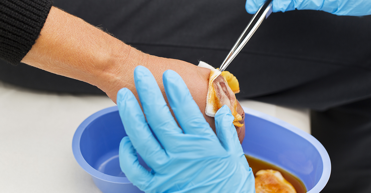
October has plenty of opportunities for a good scare. In wound care, an alarming appearance calls for a closer wound assessment.
Dark discoloration, unexpected drainage, unusual tissue, or sudden changes can certainly command a clinician’s attention. Yet appearance alone doesn’t usually tell the whole story.
The clinical challenge is to identify what you’re seeing, why it may be happening, and determine what the patient needs next.
When appearance demands a closer look
So, turn on the clinical flashlight. These seven wound scenarios highlight the importance of looking beyond the surface and teach us to interpret each finding in the context of the patient’s overall condition.
1. The wound that looks worse than expected
What are we seeing?
You remove the dressing and notice more drainage or a change in tissue appearance that wasn’t described in the previous documentation.
The immediate reaction might be: “This wound looks much worse.”
That impression deserves attention and a thorough assessment to help determine what has changed and why.
Why can it look concerning?
Changes in tissue color, drainage, or surrounding skin should naturally attract attention, particularly when they differ from the previous assessment.
Increased drainage, new or worsening redness or swelling, foul odor, increasing wound dimensions, or changes in tissue quality or color are all changes that warrant further evaluation.
What should you consider?
Compare what you’re seeing with the previous assessment and any available photographs. Review wound dimensions, tissue type, drainage, wound edges, surrounding skin, and pain, along with the patient’s overall condition.
The lesson: Let unexpected clinical findings prompt you to do a more thorough assessment.
2. The color change that raises questions
What are we seeing?
An area of skin now appears deep red, maroon, or purple and noticeably different from the surrounding skin or previous assessment.
Why can it look concerning?
A new change in skin discoloration may be clinically significant, particularly when pressure or shear is involved.
The National Pressure Injury Advisory Panel explains that underlying tissue damage leading to a deep tissue pressure injury can develop before discoloration on the skin becomes visible. Pain and temperature changes may precede changes in skin color, and the injury may evolve to reveal the full extent of deep tissue damage.
That means what you visibly see today may therefore reflect an injury that began earlier.
What should you consider?
Assess the location of pressure and shear exposure, pain, temperature, tissue consistency, skin integrity, and whether the discoloration blanches. Compare the area with surrounding tissue and previous assessments. Account for the patient’s baseline skin tone; discoloration may appear differently in dark skin. Rule out other possible causes for the change in skin color.
The lesson: Color is a clinical clue, but interpret it alongside timing and context.
3. The tissue finding that can be misunderstood
What are we seeing?
A wound bed contains tissue that looks moist, red or pink, with a bumpy cobblestone surface.
This appearance may represent healthy granulation tissue rather than deterioration.
Why can it look concerning?
Granulation tissue is part of normal wound healing but can look unfamiliar to patients or clinicians who are less experienced with wound assessment. Its textured appearance may draw attention even when it reflects healing.
Healthy tissue can also be damaged unintentionally. Protecting it from trauma during cleansing, dressing application, and dressing removal helps support continued wound healing.
WCEI’s article on granulation tissue and healing explains that healthy granulation tissue is typically red or pink and granular in appearance and that trauma can impede healing, reinforcing the need to recognize and protect this fragile tissue.
What should you consider?
Not all red tissue is healthy granulation tissue. Usually, friable tissue or tissue that bleeds readily warrants further investigation. The International Wound Infection Institute identifies bleeding, friable granulation tissue as a possible sign of local wound infection. Interpret these findings alongside pain, drainage, surrounding skin changes, and review whether cleansing techniques and dressing removal could be traumatizing newly formed tissue.
The lesson: Learn to recognize healthy versus friable granulation tissue.

4. The drainage change that gets everyone’s attention
What are we seeing?
The dressing is more saturated than the previous change, or the drainage has a new color and consistency. Or perhaps the drainage suddenly has a malodor.
Why can it look concerning?
Changes in drainage warrant reassessment, but they should be interpreted alongside other findings.
A change in wound drainage or odor becomes more concerning when it occurs alongside new or worsening swelling, redness, warmth, pain, delayed wound healing, or systemic symptoms such as fever.
For clinicians, those accompanying findings provide important context.
What should you consider?
Assess the drainage amount, color, consistency, and odor. When comparing dressing saturation, also consider the time since the last dressing change and the type and absorbency of the dressing used before making a clinical judgment.
The lesson: A changing wound deserves a fresh assessment.
5. The periwound change that shouldn’t be ignored
What are we seeing?
The wound bed itself seems relatively unchanged, but the skin around it doesn’t.
Perhaps there’s new redness, swelling, moisture damage, tenderness, pain, or breakdown.
Why can it look concerning?
The surrounding skin can reveal changes that aren’t immediately obvious when attention stays fixed on the wound bed.
Interpret these changes alongside drainage, odor, pain, healing progress, and the patient’s overall condition.
The International Skin Tear Advisory Panel recommends ongoing assessment of both the wound and surrounding skin to identify changes and guide early intervention. Periwound damage can delay healing, increase pain, and raise the risk of wound infection.
What should clinicians consider?
Look beyond the wound margin. Assess the surrounding skin’s color, temperature, edema, tenderness, moisture, and integrity. Compare changes with the patient’s previous assessment and baseline skin tone. Consider possible contributing factors.
The lesson: A complete wound assessment includes the surrounding skin.
6. The wound that isn’t progressing as expected
What are we seeing?
The wound stalls. It repeatedly breaks down. Or progress falls short of what was expected.
Why can it look concerning?
A slow-to-heal wound may indicate that something is interfering with healing, and changing the type of topical dressing alone may not address the underlying barriers.
WCEI’s article on wound bed assessment and the TIMERS framework explains that hard-to-heal wounds require a comprehensive approach that considers tissue management, inflammation or infection control, moisture balance, wound edge advancement, regeneration and repair, and social factors.
That is an important reminder: a wound doesn’t exist separately from the patient living with it.
What should you consider?
Revisit the wound’s suspected etiology and look for potential barriers to healing. Consider perfusion, pressure, edema, infection, inflammation, mobility, nutrition, medications, comorbidities, and the patient’s ability to participate in the care plan.
The lesson: When the wound doesn’t follow the expected script, reassess the script.
7. The wound that reminds us why reassessment matters
What are we seeing?
Today’s wound assessment may not look dramatically different at first glance, but its trajectory becomes clearer when current findings are compared with prior assessments.
Drainage has slowly increased. The surrounding skin has changed. Measurements have stalled. The patient’s overall health or nutritional status may also have shifted.
Individually, those changes can seem small. Together, they tell a story.
Nutrition is one example of a factor that can be easy to overlook. Changes in intake, weight, health history, or relevant laboratory values may provide important context when healing slows or stalls.
What should you consider?
Reassessment shouldn’t focus exclusively on the wound bed. Compare objective wound findings over time while also asking whether something has changed with the patient’s overall condition.
The lesson: Reassessment isn’t an extra step. It connects changes in the wound with changes in the patient.
What these wounds can teach us
The Halloween theme may be playful, but the clinical message is serious: appearance should prompt the assessment, not complete it.
A color change needs context. Drainage deserves comparison with previous findings. Surrounding skin can provide valuable clues. A wound that isn’t progressing should prompt another look at the wound, its etiology, potential barriers to healing, and the patient.
Sometimes, a wound that looks alarming may not require a major change in care. A gradual shift in drainage, stalled measurements, or a change in the surrounding skin are subtle changes that deserve attention too and become apparent only through consistent reassessment.
The real learning happens when you move beyond “What does this look like?” and ask “What is this finding telling me and what should happen next?”
Ready for Your Next Wound Care Challenge? For one week only, save $100 on eligible WCEI courses. Use code SPOOKY100 through October 31.
Explore courses

What do you think?