
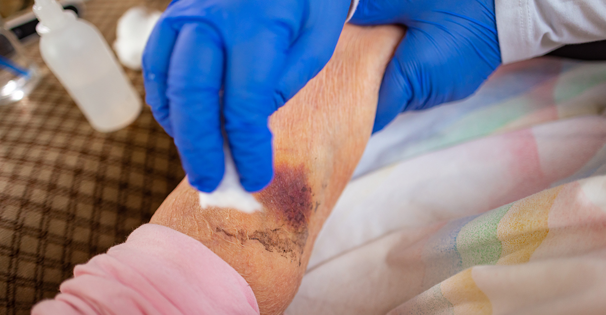
Sometimes the scariest thing in wound care isn’t the wound itself. It is what gets overlooked.
A striking wound appearance can capture your attention immediately, while subtle changes and important patient factors receive less consideration.
Assuming the wound’s etiology without adequate patient and wound assessment, overlooking changes in the surrounding skin, continuing a treatment plan without reassessing progress, or leaving gaps in documentation can all contribute to missed opportunities in patient care.
Another common pitfall is focusing so much on the wound factors that the needs of the patient living with the wound are not fully assessed.
Wound care is complex, and patients often have multiple factors that can affect healing. Developing consistent assessment habits that include a medical chart review, clear documentation, and regular reassessments can help clinicians identify concerns, reconsider the plan of care, and keep a patient-centered focus.
Five wound care mistakes to avoid
The following five mistakes can compromise assessment and an effective plan of care. Here is what to watch for and how to reduce the risk of overlooking important findings.
1. Misidentifying wound etiology
Even with the right dressing, you can still overlook the underlying problem.
Why does etiology matter?
An effective wound care plan should always begin with identifying why the wound developed.
Pressure injuries, venous ulcers, arterial ulcers, diabetic foot ulcers, moisture-associated skin damage, skin tears, and other wounds may have overlapping visual characteristics but have very different underlying causes. Appearance provides clues, but it does not establish what is driving the wound development.
For example, a lower extremity wound assessment must extend beyond the wound bed to also include perfusion status. The American Heart Association identifies slowly healing or nonhealing foot and toe wounds, cooler temperature differences between limbs, gangrene, and skin color changes among possible findings associated with peripheral artery disease (PAD). These findings would warrant further evaluation.
How to prevent this: Start by asking, What caused this tissue damage and what may be contributing to it? Consider location, pressure, perfusion, edema, neuropathy, moisture, trauma, and the patient’s medical history. Recognize that more than one factor may be involved and seek additional evaluation when the etiology is still uncertain.
2. Treating the wound without considering the whole patient
A wound may measure 3 cm by 4 cm, but size alone does not tell the whole clinical story.
Healing does not occur independently of the rest of the body. Nutrition, blood flow, glycemic management, mobility, pressure exposure, medications, comorbidities, pain, and the patient’s ability to participate in the plan of care can influence both healing and the feasibility of treatment recommendations.
For patients with diabetes, the International Working Group on the Diabetic Foot recommends a comprehensive patient assessment that includes ulcer characteristics, peripheral artery disease, infection, neuropathy, foot deformity, mobility, comorbidities, and social circumstances.
A whole-person approach is essential to effective wound management.
How to prevent this: Before thinking you need to choose another dressing option, ask whether another factor could be interfering with healing. The next step may involve addressing pressure, perfusion, nutrition, glycemic management, mobility, or other patient-specific concerns.

3. Missing changes in the surrounding skin
It’s easy to develop tunnel vision when the wound bed is the most obvious part of the assessment.
But the surrounding skin may reveal what the wound bed does not.
Changes in skin color, texture, temperature, moisture, swelling, or integrity may provide valuable information about what is happening around the wound.
Wounds International’s expert consensus, Best Practice Recommendations for Prevention and Management of Periwound Skin Complications, emphasizes that the periwound is an essential part of wound assessment. Changes such as maceration, inflammation, erosion, or skin stripping may signal complications that can delay healing, increase pain, and raise the risk of infection. Early identification of periwound changes allows clinicians to address contributing factors and protect skin integrity before further damage occurs.
How to prevent this: Don’t stop at the wound margin. Make periwound skin assessment part of every wound evaluation. Best practice is to assess the skin circumferentially at least 4 cm beyond the wound edge, extending the assessment farther when abnormalities are present and including all skin beneath the dressing.
4. Assuming one sign means infection
Wound odor, purulent drainage, increased pain, redness and inflammation, and wound debris all deserve attention — but no single sign confirms infection. You should evaluate findings collectively, compare them with the patient’s baseline, and consider whether they indicate a meaningful change in the wound or patient’s overall condition.
The Centers for Disease Control and Prevention’s surgical site infection basics list redness and pain around a surgical site, cloudy drainage, and fever among signs and symptoms of surgical site infection. These findings should prompt further assessment along with interpretation of all findings.
Odor, for example, may result from devitalized tissue, excess exudate, or a dressing left in place too long; it should prompt assessment rather than an immediate conclusion of infection.
How to prevent this: Don’t rely on just a single wound characteristic. Look for other changes and patterns. Consider the wound bed appearance, type and amount of drainage, pain, surrounding tissue, temperature, systemic findings, and the patient’s overall clinical status, and follow facility protocols for escalation when infection is suspected.
5. Documenting the wound, but not its whole story
“Wound looks better.” That sounds encouraging, but it leaves the next clinician with a question: Better how?
Objective wound documentation makes it possible to understand what was observed, compare assessments, and track changes over time.
The American Nurses Association identifies clear, accurate, and accessible documentation as an essential element of safe, quality, evidence-based nursing practice.
For wound care, meaningful documentation may include measurements, tissue characteristics, wound edges, drainage, surrounding skin, pain, interventions, and the patient’s response, as appropriate to the wound, setting, and organizational requirements. Using consistent wound description terms can also help clinicians communicate findings clearly.
How to prevent this: Replace vague descriptions with observable findings. Consistency matters because each assessment should provide a clear point of comparison over time.
The mistake behind many mistakes: failing to reassess
Yesterday’s treatment plan may have made perfect sense yesterday. That doesn’t mean it should continue indefinitely without reassessment.
Reassess at regular intervals and whenever the wound, the patient’s condition, or the response to treatment changes or raises concern.
Ask yourself, Is the suspected etiology still supported by the findings? Has perfusion changed? Is pressure adequately managed? Has edema increased? Are there new infection concerns? Has the patient’s overall health or ability to follow the care plan changed?
Reassessment is not an admission that the plan failed. It is how clinicians confirm that the plan still reflects the wound, the patient, and the factors affecting healing.
Sometimes the most useful question isn’t “What dressing should I use?”
It’s “What might I be missing?”
Build Your Wound Care Knowledge With WCEI. Save $100 on eligible WCEI courses from October 25 through October 31, 2026, with code SPOOKY100.
Explore courses

What do you think?