Key Takeaways
Full thickness wounds penetrate through the epidermis and dermis into subcutaneous tissue and possibly beyond, requiring targeted interventions due to their complexity and potential severity. Categorization systems like those from the NPIAP, Wagner, and ISTAP help classify wounds and guide appropriate treatment strategies, but when wounds don’t fit neatly into these categories, understanding tissue depth is crucial for determining care. Effective treatment involves managing drainage, preventing infection, and fostering healing, with patient education and interdisciplinary referrals playing vital roles in successful wound management.
One important step in wound management is understanding and knowing just how deep the damage goes in a wound. Full thickness wounds aren’t just skin-deep, they’re a call for targeted interventions.
Most wounds are categorized as superficial, partial thickness, or full thickness. But here’s where it gets interesting: Certain types of wounds have their own specialized classification systems that fit within these general depth categories.
You’ve probably heard of the National Pressure Injury Advisory Panel (NPIAP) staging system for pressure injuries or the Wagner scale for grading diabetic foot ulcers. Maybe you’re familiar with the International Skin Tear Advisory Panel (ISTAP) system for skin tears, too. These tools help us speak the same language when it comes to wound severity and guide us in choosing the right treatment approach.
But some wounds don’t fall neatly into these categories. How do you approach those?
That’s where understanding the level of tissue loss is a must. When wounds fall outside those specific subtypes, we assess them by depth, because the deeper the tissue damage, the more complex the healing process and the more tailored treatment needs to be. Knowing how to identify and classify these wounds isn’t just academic, it’s the foundation of effective care.
Understanding wound depth classification
Let’s take a closer look at wound depth classifications. Each of the following levels of depth reveals important clues about the wound’s severity and what it needs to heal effectively:
- Superficial wounds: The tissue injury is usually limited to the epidermis but may extend to the very outer Examples include small scratches or skin abrasions.
- Partial thickness wounds: The tissue injury extends through the epidermis and into the dermis without going into the subcutaneous tissue. Examples include second-degree burns and deeper trauma
- Full thickness wounds: The tissue injury extends through the epidermis and dermis into the subcutaneous tissue, making it the most complex and potentially serious of the three classifications.
Measuring a wound’s depth to monitor for healing requires a sterile foam-tipped applicator to be applied to the deepest part of the wound bed, perpendicular to the skin’s surface. The point where the applicator meets the opening of the wound is the depth.
This depth is noted, usually by placing the fingertips at the point where the wound meets the skin’s surface, by comparing this measurement to a ruler. Documenting wound depth with each assessment will help determine how the wound is healing.
Another method to help determine the depth is visual inspection. Do you see fat, muscle, or fascia in the wound bed? If so, you have a full-thickness wound
Full thickness wounds: Definition and causes
A full thickness wound extends through the epidermis and dermis into the subcutaneous tissue below. However, it may extend further through the subcutaneous tissue and into the supporting structures below, such as the fascia, muscle, tendon, cartilage, bone, and ligament.
Remember, subcutaneous tissue (also known as the hypodermis) is primarily composed of fat cells. If you’re seeing a soft white or pale-yellow tissue in the wound bed, you’re looking at the subcutaneous layer, which makes the wound full thickness. These wounds are typically caused by pressure, friction, shearing, ischemia, burns, surgery, or trauma.
Here are some common examples of wounds that may be classified as full thickness:
- Pressure injuries
- Diabetic foot ulcers
- Venous ulcers
- Arterial ulcers
- Trauma wounds other than skin tears
- Autoimmune ulcers
- Radiation injuries
- Surgical wounds
- Abscesses
- Burns

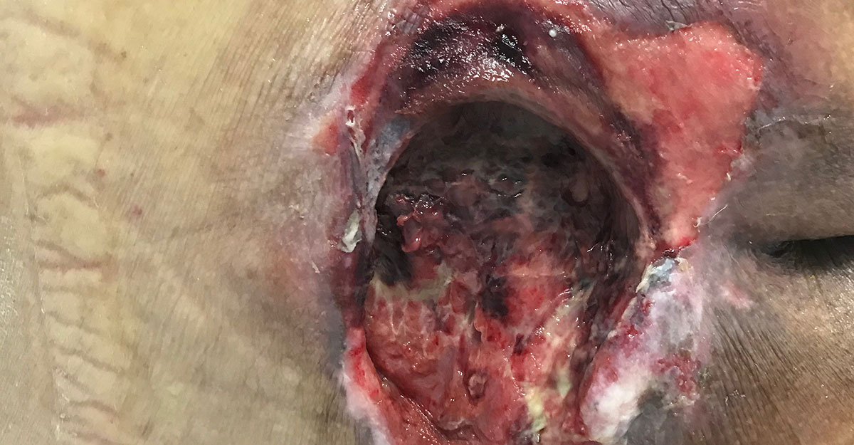
Characteristics of full thickness wounds
Full thickness wounds may have a mixture of granulation tissue and necrotic tissue, including slough and eschar. Due to tissue loss, full thickness wounds usually have some amount of drainage.
The drainage may be serous or serosanguineous; however, tan or green purulent drainage indicates possible infection. Other signs of infection in full thickness wounds include induration, increased drainage, warmth, redness, and odor.
Full thickness wounds may be painful, especially if infected, or they may have very little pain, due to nerve damage.
Treatment strategies
Controlling drainage in these wounds is important to the periwound skin’s integrity. It also prevents infection and creates an optimal wound environment. So choosing dressings that encourage tissue growth and control drainage, such as alginate or collagen dressings, is a must.
Also, consider treatments such as growth factors to encourage granulation tissue growth. Some full thickness wounds with adequate blood supply may benefit from negative pressure wound therapy or skin substitutes.
However, serial debridement may also be necessary to allow new growth of granulation tissue. Removing necrotic tissue decreases the risk of infection and helps move the wound out of the inflammatory phase.
Healing process and prognosis
Full thickness wounds will move through the stages of healing (hemostasis, inflammation, proliferation, and remodeling). And the timeframe for healing depends on the wound’s movement through these stages.
Often, wounds will stall in the inflammatory phase until infection has been treated and wound conditions, such as maintaining a moist, warm environment, have improved. Like all wounds, ensuring adequate blood flow and instructing the patient on nutritional support also promotes effective healing.
A progressing wound will have signs of improved tissue quality and measurable signs of healing. Complications should be identified and remedied as soon as possible to prevent stalling and regression. Treating infection, ensuring adequate blood flow, managing venous insufficiencies, relieving pressure, friction, or shearing forces, and controlling drainage will help the wound progress.
Considerations and patient education
Pain management is a key concern in wound care. Both acute and chronic pain should be addressed through safe, effective methods, including medication as prescribed and edema control. Educate patients on these approaches to empower their self-care.
Refer patients with compromised arterial flow to a vascular surgeon. Recurrent or resistant infections may warrant referral to an infectious disease specialist. Primary care providers and endocrinologists play essential roles in managing medications and chronic conditions like diabetes. Other helpful referrals may include general surgery, orthotics, podiatry, and foot and ankle specialists.
Patient education is critical, as most care occurs outside the clinic. Teach patients proper dressing change techniques and signs of infection. Have them demonstrate the process when necessary to ensure competence. For added support, consider involving a home health nurse.
Reinforce education on edema management, nutrition, offloading, orthotic compliance, and following through with referrals. Each of these elements is vital to successful healing.
Final thoughts on full thickness wounds
Full thickness wounds represent a large portion of wounds requiring specialized treatment. Knowing how to identify and treat them can make a difference in your patients’ lives. Being a part of those life-changing stories is what keeps us pushing on.
Elevate your expertise with education from WCEI!
Get StartedWhat do you think?